HPLC-MS (also written LC-MS) joins a liquid chromatograph to a mass spectrometer, so each compound separated by the column is weighed as it comes off. On a peptide COA it is the usual way to confirm identity: the lab compares the mass it observed with the mass the peptide's sequence predicts, and a match is strong evidence that the main peak is the right molecule.
This note explains how a mass spectrometer weighs a peptide, how to read the masses on a certificate, and what mass spectrometry can't tell you.
What is HPLC-MS?
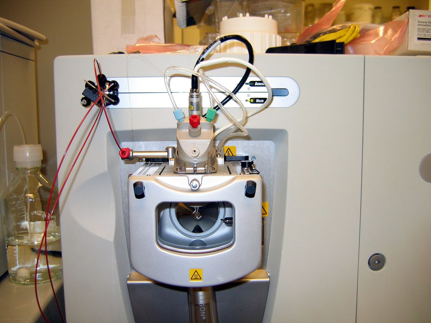
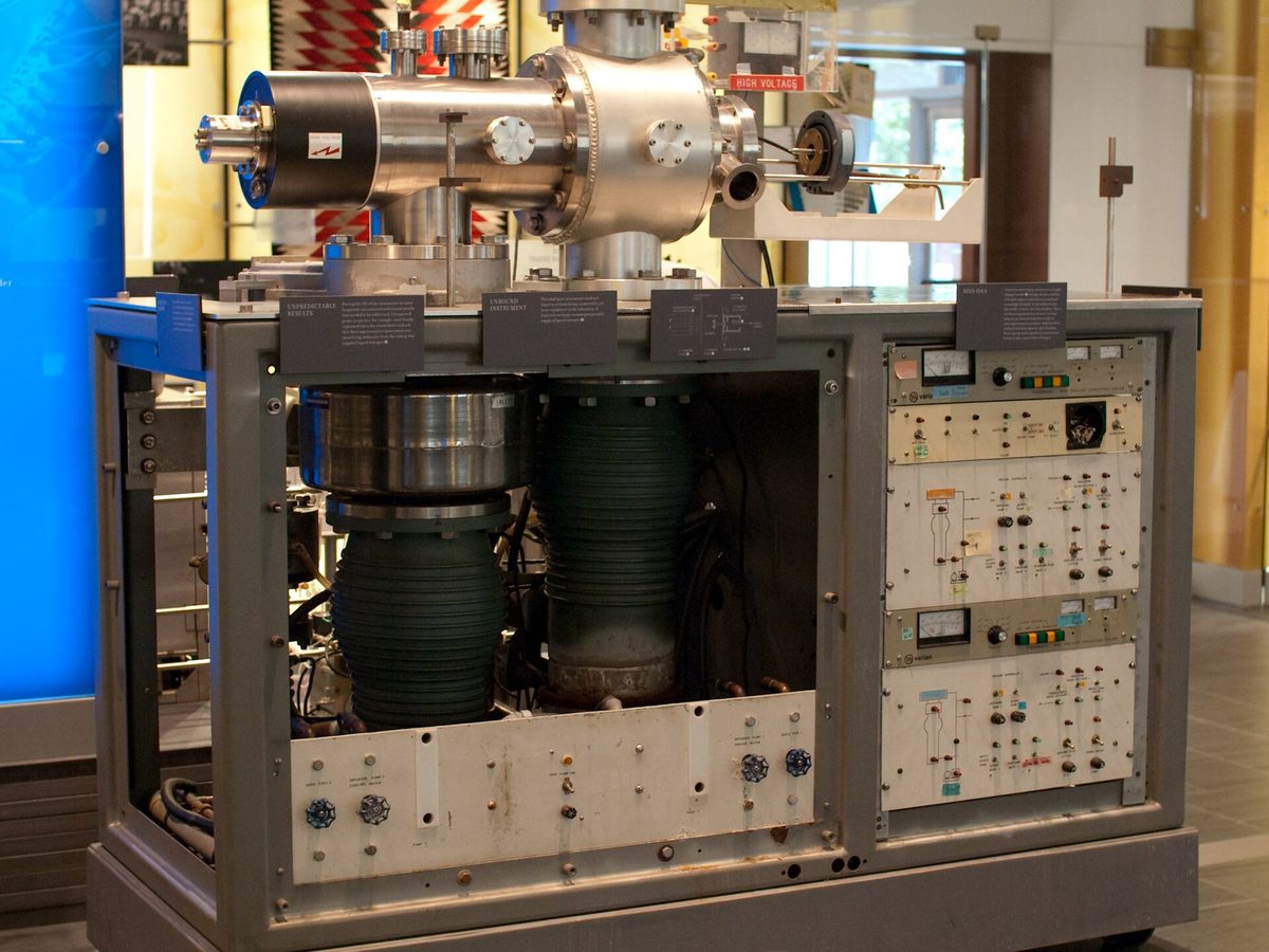
In HPLC-MS the column does the separating and the mass spectrometer does the identifying. Harvey's analytical chemistry text describes the key engineering problem: the liquid mobile phase is incompatible with the vacuum a mass spectrometer needs, so the column effluent enters the ion source through an interface that removes most of the mobile phase. What reaches the analyser is a stream of charged molecules.
The mass spectrometer measures the mass-to-charge ratio (m/z) of those ions. A UV detector can tell you that something left the column at 12.8 minutes; a mass spectrometer can tell you that what left at 12.8 minutes has the mass of your peptide.

How electrospray turns a peptide into ions
Most peptide identity work uses electrospray ionisation (ESI). In the method John B. Fenn published in 1988, the Nobel Prize committee explains, charged droplets of protein solution are produced which shrink as the water evaporates, leaving free ions whose masses can then be measured. Fenn and Koichi Tanaka shared half of the 2002 Nobel Prize in Chemistry for their development of soft desorption ionisation methods for mass spectrometric analyses of biological macromolecules.
ESI is called a soft ionisation method because it leaves the molecule largely intact, so its molecular weight information survives. Large molecules such as peptides readily pick up multiple protons, forming ions of the form [M + zH]z+, where z is the number of protons added. One peptide therefore appears as several peaks, one for each charge state.

Reading a mass spectrum on a COA
Fig. 3 shows an illustrative spectrum for a fictional peptide with an expected mass of 1,418.70 Da. The same molecule appears three times: once carrying one proton, once carrying two and once carrying three.
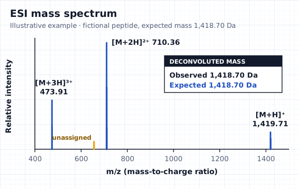
The arithmetic is simple enough to check by hand. A proton weighs about 1.007 Da, so for a charge state z, the neutral mass M is the m/z value multiplied by z, minus z protons:
| Ion | m/z | Calculation | Neutral mass (Da) |
|---|---|---|---|
| [M+H]⁺ | 1,419.71 | 1,419.71 × 1 − 1.007 | 1,418.70 |
| [M+2H]²⁺ | 710.36 | 710.36 × 2 − 2.014 | 1,418.71 |
| [M+3H]³⁺ | 473.91 | 473.91 × 3 − 3.021 | 1,418.71 |
Software usually does this step, called deconvolution, and reports a single observed mass. A clear identity result on a COA therefore has three parts: the observed mass, the expected mass for the sequence, and the spectrum or at least the main ions behind it. The report should also say whether it quotes monoisotopic or average mass, because the two values differ for the same peptide.
What "identity confirmed" should look like
"Identity: conforms" with nothing else is a conclusion without evidence. A useful identity line reads something like "observed 1,418.70 Da, expected 1,418.70 Da, LC-ESI-MS", with the spectrum attached. Peptide Testing Australia's guide to how purity and identity sit side by side on a test result makes the same point: the observed mass has to match the expected mass, regardless of how pure the sample looks.
What mass spectrometry can't tell you
- Same-mass isomers. A peptide with one amino acid in the wrong mirror-image form (a D instead of an L residue) has the same mass as the correct one. The EMA's guideline on synthetic peptides notes that controlling diastereomers may require the development of specific methods.
- Sequence order. Two peptides with the same amino acids in a different order share a mass. The EMA guideline notes that variants of mass spectrometry can determine molecular mass and confirm the amino acid sequence; fragmentation (tandem MS) does the second job, and a single intact mass can't.
- Purity. A mass spectrum shows which ions are present, but signal strength varies from compound to compound, so it isn't a substitute for an HPLC purity figure.
- Content and endotoxin. Like HPLC, mass spectrometry doesn't measure peptide content by weight or detect endotoxin.
This is why the EMA guideline recommends at least two orthogonal methods for identification, listing mass, relative retention time, LC-MS, peptide mapping, bioactivity, amino acid analysis and NMR among the acceptable options, and why it asks applicants to show the identification tests can unambiguously confirm the peptide's sequence.
Why identity and purity need each other
Identity and purity answer different questions, and a COA needs both to say much about a vial. A sample can be 99% pure by HPLC and still be the wrong peptide, because HPLC measures how much of the signal is one compound, not which compound it is. A sample can also have the right mass and be only 70% pure, with the rest made up of closely related impurities that the main mass doesn't reveal.
Reading the two results together is what makes them useful. The mass spectrum should come from the same sample, ideally the same run, as the purity figure. When an LC-MS run is used, the lab can show that the main UV peak and the expected mass appear at the same retention time, which ties the purity number to the right molecule. A report that gives a purity figure from one lab and an identity claim from another, or from a different batch, hasn't linked the two.
Questions to ask about an identity result
- What was the expected mass, and is it monoisotopic or average?
- Which ions were observed, and can I see the spectrum?
- Was the mass spectrum run on the same sample as the HPLC purity?
- Were any unassigned masses seen, and at what level?
Questions
What is HPLC-MS?
HPLC-MS, or LC-MS, is a liquid chromatograph connected to a mass spectrometer. The column separates the sample and the mass spectrometer measures the mass-to-charge ratio of each compound as it leaves, which is how a peptide's identity is usually confirmed.
What is the difference between HPLC and mass spectrometry?
HPLC separates compounds and, with a UV detector, measures how much signal each gives, which is used for purity. Mass spectrometry measures molecular mass, which is used for identity. A COA normally needs both.
Why does my COA show several masses for one peptide?
Electrospray ionisation adds different numbers of protons to the same molecule, so one peptide appears at several m/z values. Each should calculate back to the same neutral mass.
Can mass spectrometry prove a peptide is pure?
No. It confirms which ions are present but doesn't give a reliable percentage purity, and it can't separate same-mass isomers. Purity comes from HPLC; identity is strongest with at least two different methods.